Once the breast cancer and the armpit lymph glands have been removed, they are then examined under the microscope by a specialist called a pathologist. The pathologist will determine the following:
- Exactly what type of breast cancer it is?
- How big is the cancer?
- Has the cancer been completely removed? Are the surgical margins clean?
- Do the lymph glands contain cancer?
- Does the cancer have hormone receptors?
- Does the cancer have the growth protein Her2?
The pathologist will also decide the severity of the cancer according to its appearance under the microscope and how much it has spread. In practice, the cancer is assessed (or staged).
When is it Advisable to Have a Mastectomy Rather than a Lumpectomy?
In certain situations, your breast specialist may advise you to have complete removal of the breast (mastectomy), rather than removal of the cancer lump (lumpectomy). Such situations include the following:
- If the cancer is recurrent and you previously had radiotherapy.
- If the tumour lies in a central location behind or close to the nipple.
- If there is more than one cancer in the same breast.
- If you carry a specific gene mutation.
- If the cancer is very large in relation to the size of the breast (more than 4 cm wide).
- If radiotherapy is contraindicated
- If the patient specifically requests a mastectomy.
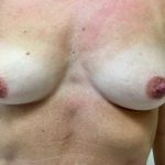
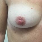
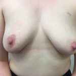
The aesthetic outcome of lumpectomy and radiation therapy for right sided breast cancer through a scar around the right nipple
Possible Problems Following Breast Surgery
Most patients do not develop any problems after surgery. However, the following problems can occur in some cases.
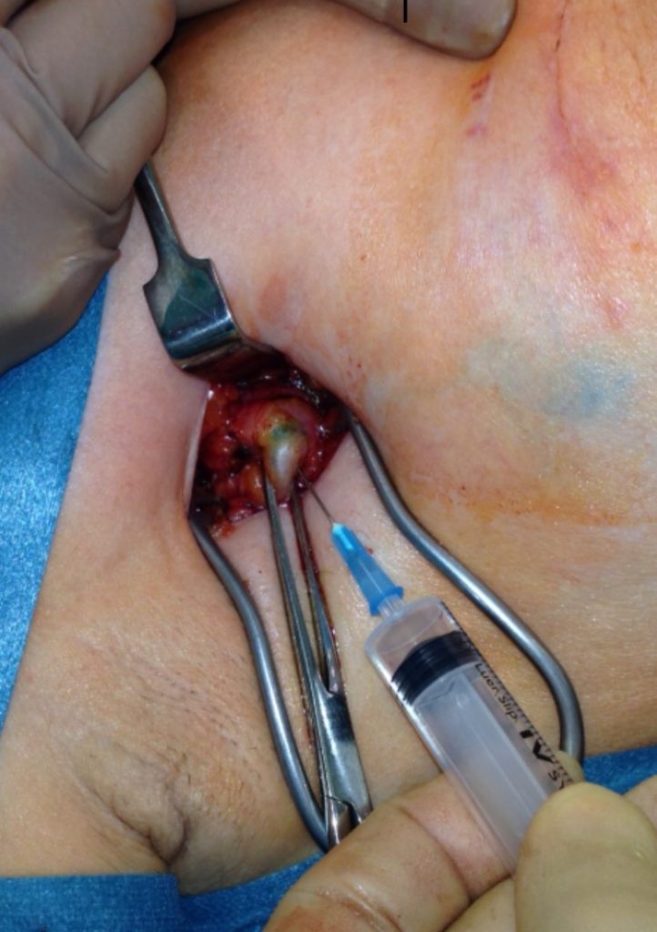
Wound Infection
This problem affects around 2 out of every 100 patients. It usually occurs a few days after surgery, causing pain and redness of the wound. Infections can be treated easily using antibiotics. Occasionally, a collection of pus can develop under the wound, called an abscess. If this happens, then the wound stitches are removed allowing the pus to be drained.
Blood Collection in the Wound (Haematoma)
It usually appears within 24 hours after surgery, causing swelling and pain in the wound. Only very large haematomas require treatment.
Fluid Collection in the Armpit (Seroma)
During removal of the lymph glands in the armpit, the lymph channels are also cut. This causes lymphatic fluid to collect instead of being drained away; it accumulates under the skin of the armpit wound, forming a swelling called a seroma. The leakage of lymph fluid will eventually stop and the swelling will disappear. However, if the seroma is causing significant discomfort, it can be drained using a needle and syringe.
Swelling of the Arm (Lymphoedema)
The lymphatic channels of the arm transport lymph fluid back into the blood circulation. These channels pass through the lymph glands in the armpit. Removal or damage to these armpit glands can result in accumulation of lymph fluid in the arm, causing severe swelling called lymphoedema. It occurs in around 5% of patients undergoing axillary dissection. The figure is much higher if the armpit is also treated with radiation. This complication is rare after axillary node sampling or sentinel node biopsy procedure.
Unfortunately, there is no simple surgical solution to lymphoedema. Treatment involves physiotherapy, elevation of the arm, wearing compression sleeves and using antibiotics to treat early infections. Recent evidence suggests that microsurgery to connect the lymphatics to the veins is effective in reducing lymphoedema.
Shoulder Stiffness
Reduced shoulder mobility is a recognised problem after breast cancer surgery. A physiotherapist will teach you specific exercises to improve the problems. Occasionally scar bands develop which restrict shoulder movements. This can also be improved with physiotherapy. Release of these bands using surgery or laser is sometimes required. This complication is uncommon after sentinal node biopsy.
Burning Pain and/or Numbness in the Inner Side of the Upper Arm
This often occurs after axillary surgery to remove the lymph nodes from the armpit. This complication is due to the cutting of the nerve supplying the area during surgery. The symptoms tend to improve with time. This complication is uncommon after sentinal node biopsy.
‹‹Previous Next››
